















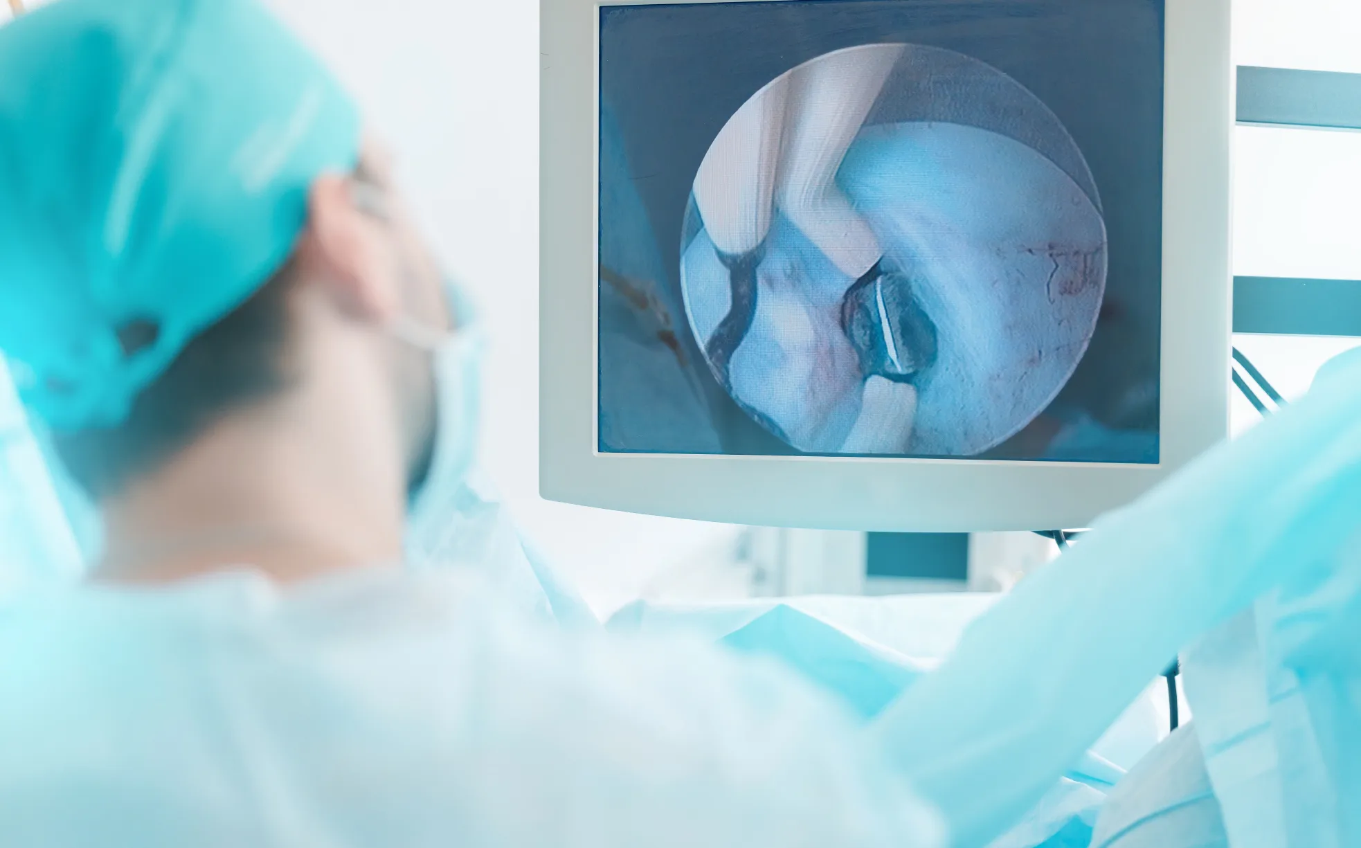
Colonoscopy plays an important role in colorectal cancer prevention by detecting and removing precancerous polyps. However, not all polyps carry the same risk of progressing to cancer, which makes careful assessment during the procedure important.
Some adenomas have features associated with a higher risk of advanced changes or future colorectal cancer. Identifying these lesions helps guide appropriate management and follow-up after colonoscopy.
How Does a Colonoscopy Doctor Identify High-Risk Adenomas?
During colonoscopy, the doctor assesses the characteristics of any polyps identified to estimate their nature and look for features associated with more advanced changes. The final risk assessment also takes into account pathology findings after the polyp is removed. This assessment can help guide how the lesion is managed during the procedure and whether further evaluation is needed.
Assessing the Polyp's Appearance
The doctor assesses features such as the polyp's size, shape, colour, surface pattern and borders. Larger adenomas are generally more likely to contain advanced changes, although size alone does not determine risk. The overall appearance helps the doctor assess the lesion and decide how it should be managed during the procedure.
Using Narrow-Band Imaging
Narrow-band imaging (NBI) enhances the appearance of blood vessels and surface patterns within the lining of the colon. This can help the doctor characterise a polyp more closely and identify features associated with adenomatous or more advanced changes during the procedure.
Recognising Subtle Serrated Lesions
Sessile serrated lesions are often flat, pale and covered by a thin layer of mucus, making them more difficult to detect than conventional adenomas. Although they are a distinct type of precancerous colorectal lesion, they can also progress towards colorectal cancer. Careful inspection is therefore important, particularly because these lesions commonly occur in the proximal colon.
How Do Higher-Risk Adenoma Findings Affect Management?
Once an adenoma is identified, its characteristics help determine the most appropriate next steps. Management may differ depending on the lesion’s size, location, appearance and complexity.
Determining the Most Appropriate Removal Method
Many adenomas can be removed during the colonoscopy. Larger or more complex lesions may require more advanced removal techniques to achieve complete excision while managing the risk of complications. The approach depends on the lesion’s size, location, morphology and whether there are features suspicious for early cancer.
When Advanced Endoscopic or Surgical Treatment May Be Needed
Some adenomas may be too large or technically complex for routine removal, while others may have features suspicious for early colorectal cancer. In these cases, advanced endoscopic removal or surgical assessment may be recommended depending on the characteristics of the lesion.
Planning Appropriate Surveillance
Patients with higher-risk adenoma findings may require follow-up colonoscopy at a shorter interval than those with low-risk findings. The recommended surveillance interval depends on factors such as the number and size of adenomas, their pathology and whether they were completely removed.

Why Does Experience Matter When Performing Colonoscopy?
The quality of a colonoscopy depends not only on the imaging technology used but also on how carefully the examination is performed and how findings are interpreted. Experience can be particularly important for:
- Recognising subtle lesions: Some polyps are flat, small or difficult to distinguish from the surrounding bowel lining and may require careful inspection to identify.
- Assessing lesions during the procedure: The doctor considers a polyp’s appearance alongside enhanced imaging where appropriate to determine how it should be managed.
- Choosing an appropriate removal approach: The size, location and complexity of a lesion can influence whether it can be removed during the procedure or requires advanced endoscopic or surgical management.
- Planning appropriate follow-up: Findings during colonoscopy, together with subsequent pathology results, help determine whether and when surveillance colonoscopy may be needed.
Assessment and Management of Adenomas at Colorectal Practice
A colonoscopy is more than a screening test to find polyps. It also involves carefully assessing each lesion for features that may influence how it is removed and whether further evaluation is needed. Pathology findings after removal then help guide subsequent treatment and surveillance. Technologies such as narrow-band imaging, combined with the experience of a skilled colonoscopy doctor and informed clinical decision-making.
At Colorectal Practice, we are committed to delivering personalised care through the expertise of our experienced surgeons. Our clinical team is led by our medical director and senior consultant colorectal surgeon, Dr Dennis Koh, alongside Dr Sharon Koh and Dr Pauleon Tan, our other senior consultant colorectal surgeons, who work closely with relevant healthcare professionals to ensure treatment recommendations remain patient-centred and evidence-based. Schedule a consultation and take the next step towards appropriate colorectal care.
Frequently Asked Questions (FAQs) About Adenomas
Can a doctor tell if a polyp is high risk during a colonoscopy?
A colonoscopy doctor can assess features such as a polyp’s size, shape, surface pattern and blood vessel appearance to estimate whether it may have higher-risk characteristics. Narrow-band imaging (NBI) can provide additional detail during this assessment, while examination of removed tissue by a pathologist provides information about its microscopic features.
Are all adenomas considered precancerous?
Adenomas are considered precancerous because they have the potential to develop into colorectal cancer, although most do not progress to cancer. The level of risk varies according to factors such as the adenoma’s size and microscopic features, including the degree of dysplasia.
What is a sessile serrated lesion?
A sessile serrated lesion is a type of precancerous polyp that is often flat and difficult to detect during colonoscopy. It differs from a conventional adenoma but can also progress towards colorectal cancer, which makes careful detection and complete removal important.
If a high-risk adenoma is removed, will I need another colonoscopy?
In most cases, follow-up colonoscopy will be recommended after higher-risk adenoma findings. The timing depends on factors such as the number and size of adenomas, their pathology, whether they were completely removed and other relevant colonoscopy findings.
Does narrow-band imaging replace pathology testing?
No. Narrow-band imaging helps the doctor assess a polyp’s characteristic during colonoscopy, but it does not replace histopathological examination when removed tissue is sent for analysis. Pathology provides information about the type of lesion and its microscopic features.
Meet Our Colorectal Specialists in Singapore